
Please use this identifier to cite or link to this item:
http://hdl.handle.net/11054/1580Full metadata record
| DC Field | Value | Language |
|---|---|---|
| dc.contributor | Amir, N. S. S. | en_US |
| dc.contributor | Zhe Kang, Law | en_US |
| dc.contributor | Mukari, S. A. | en_US |
| dc.contributor | Sahathevan, Ramesh | en_US |
| dc.contributor | Chellappan, K. | en_US |
| dc.date.accessioned | 2020-08-10T09:45:42Z | - |
| dc.date.available | 2020-08-10T09:45:42Z | - |
| dc.date.issued | 2020 | - |
| dc.identifier.govdoc | 01507 | en_US |
| dc.identifier.uri | http://hdl.handle.net/11054/1580 | - |
| dc.description.abstract | A critical step in detection of primary intracerebral haemorrhage (ICH) is an accurate assessment of computed tomography (CT) brain images. The correct diagnosis relies on imaging modality and quality of acquired images. The authors present an enhancement algorithm which can improve the clarity of edges on CT images. About 40 samples of CT brain images with final diagnosis of primary ICH were obtained from the UKM Medical Centre in Digital Imaging and Communication in Medicine format. The images resized from 512 × 512 to 256 × 256 pixel resolution to reduce processing time. This Letter comprises of two main sections; the first is denoising using Wiener filter, non-local means and wavelet; the second section focuses on image enhancement using a modified unsharp masking (UM) algorithm to improve the visualisation of ICH. The combined approach of Wiener filter and modified UM algorithm outperforms other combinations with average values of mean square error, peak signal-to-noise ratio, variance and structural similarity index of 2.89, 31.72, 0.12 and 0.98, respectively. The reliability of proposed algorithm was evaluated by three blinded assessors which achieved a median score of 65%. This approach provides reliable validation for the proposed algorithm which has potential in improving image analysis. | en_US |
| dc.description.provenance | Submitted by Gemma Siemensma (gemmas@bhs.org.au) on 2020-08-03T01:19:07Z No. of bitstreams: 0 | en |
| dc.description.provenance | Approved for entry into archive by Gemma Siemensma (gemmas@bhs.org.au) on 2020-08-10T09:45:42Z (GMT) No. of bitstreams: 1 HTL.2018.5003.pdf: 357006 bytes, checksum: 73f37599943c649fa1570921c60c912d (MD5) | en |
| dc.description.provenance | Made available in DSpace on 2020-08-10T09:45:42Z (GMT). No. of bitstreams: 1 HTL.2018.5003.pdf: 357006 bytes, checksum: 73f37599943c649fa1570921c60c912d (MD5) Previous issue date: 2020 | en |
| dc.title | CT brain image advancement for ICH diagnosis. | en_US |
| dc.type | Journal Article | en_US |
| dc.type.specified | Article | en_US |
| dc.bibliographicCitation.title | Healthcare Technology Letters | en_US |
| dc.bibliographicCitation.volume | 7 | en_US |
| dc.bibliographicCitation.issue | 1 | en_US |
| dc.bibliographicCitation.stpage | 1 | en_US |
| dc.bibliographicCitation.endpage | 6 | en_US |
| dc.subject.healththesaurus | COMPUTER TOMOGRAPHY | en_US |
| dc.subject.healththesaurus | CT | en_US |
| dc.subject.healththesaurus | INTRACEREBRAL HAEMORRHAGE | en_US |
| dc.subject.healththesaurus | STROKE | en_US |
| dc.subject.healththesaurus | ALGORITHM | en_US |
| dc.identifier.doi | http://doi.org/10.1049/htl.2018.5003 | en_US |
| Appears in Collections: | Research Output | |
Files in This Item:
| File | Description | Size | Format | |
|---|---|---|---|---|
| HTL.2018.5003.pdf | This is an open access article published by the IET under the Creative Commons Attribution License | 348.64 kB | Adobe PDF | 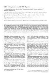 View/Open |
Items in DSpace are protected by copyright, with all rights reserved, unless otherwise indicated.